Featured Videos
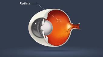
A retinal detachment is a separation of the inner lining of the eye. This causes a hole in the retina, and through that hole liquid causes a separation. The eye has many structures, including the cornea in the front of the eye, the lens in the middle, and the retina, which lines the inside of the structure of the wall. Generally, a retinal detachment starts in the peripheral part of the retina, and can extend towards the central area.
Retinal Detachment Symptoms
A retinal detachment requires immediate medical care, as it can lead to the loss of central vision. Retinal detachment causes include being highly myopic (nearsighted), patients who have experienced trauma to the eye, and patients who have undergone cataract surgery. Approximately one percent of patients who have had cataract surgery will develop a retinal detachment, usually during the year or two after the cataract surgery. Retinal detachment symptoms include flashes of light, floaters and loss of peripheral vision. However, some patients don’t notice any symptoms, or only notice an issue when they lose vision. A retinal detachment almost always requires surgery to put the retina back in place.
Retinal Detachment Treatment
There are a few different retinal detachment surgeries your ophthalmologist may suggest:
• Scleral buckle : The scleral buckle procedure is performed in the operating room with local or general anesthesia. The ophthalmologist secures a buckle to the wall of the eye, creating a scar to ensure that the retinal tear stays sealed. Typically, the eye surgeon also drains the sub-retinal fluid.
• Pneumatic retinopexy: The ophthalmologist freezes the eye with anesthetic, injects a gas bubble into the eye and creates a tear adhesion with cryotherapy or laser. Your head position will be restricted after the procedure to keep the gas bubble in place.
• Vitrectomy : This is the most common way of repairing a retinal detachment. During this procedure, the ophthalmologist removes the vitreous gel that is pulling on the retina, usually replacing it with a gas bubble. If a gas bubble is used, your ophthalmologist may recommend that you keep your head in special positions for a time.
Your ophthalmologist will determine the best retinal detachment treatment for your situation. A patient’s age and their previous ocular history, including history of surgery, do have implications in terms of what type of repair your vitreoretinal surgeon might choose to use. For example, a younger patient might be more likely to have a scleral buckle procedure. Vitrectomy causes cataract, so a patient who is older or a patient who has already had cataract surgery performed would be more likely to have a vitrectomy operation.
Talk to your eye doctor if you'd like more information on retinal detachment.
Healthy eyes depend on regular visits to your optometrist for eye exams, and if necessary, an ophthalmologist for certain eye conditions and surgeries like diabetic retinopathy . You can also protect your eyesight with proper nutrition, eating foods that contain the right vitamins. Local Optometrists may prescribe eyeglasses and contact lenses, provide laser eye surgery consultations, and test for diseases. Local Ophthalmologist can help with many facts of eye diseases. Getting a referral from your optometrist to a local ophthalmologist is crucial to eye care.

